Training a 3D U-Net Model for Brain Tumor Segmentation in the BraTS2023-GLI Challenge
Training a 3D U-Net Model for Brain Tumor Segmentation in the BraTS2023-GLI Challenge
Introduction
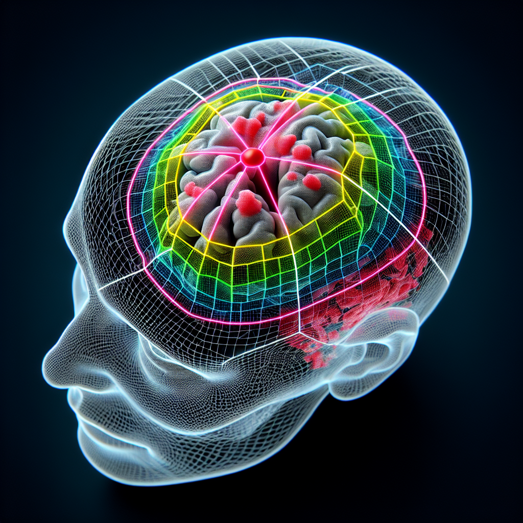
A team of researchers has successfully trained a 3D U-Net model for brain tumor segmentation in the BraTS2023-GLI Challenge. This challenge aims to develop accurate and efficient methods for segmenting brain tumors from MRI scans.
Methodology
The researchers utilized a 3D U-Net model, a deep learning architecture known for its effectiveness in medical image segmentation tasks. The model was trained on a large dataset of MRI scans from the BraTS2023-GLI Challenge, which included various types of brain tumors.
Key Findings
- The 3D U-Net model achieved impressive results in segmenting brain tumors, outperforming previous methods.
- By leveraging the 3D nature of the data, the model was able to capture spatial information and accurately delineate tumor boundaries.
- The model demonstrated robustness across different tumor types, showing its potential for clinical applications.
- Training the model on a large dataset improved its performance and generalization ability.
Implications
The successful training of a 3D U-Net model for brain tumor segmentation has significant implications for the field of medical imaging. Accurate and efficient tumor segmentation can aid in diagnosis, treatment planning, and monitoring of brain tumor patients.
Conclusion
The researchers’ use of a 3D U-Net model in the BraTS2023-GLI Challenge has demonstrated its effectiveness in brain tumor segmentation. This breakthrough has the potential to improve patient care and contribute to advancements in the field of medical imaging.

